General Information
- A congenital dermal sinus is a scaly, multi-layered channel of tissue found along the body’s midline anywhere between the nasal bridge and the tailbone.
- The tract may end just below the skin surface or may extend to portions of the spinal cord, skull base or nasal cavity.
Symptoms
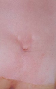
- A spinal dermal sinus may appear as a dimple or a sinus (open tract), with or without hairs, usually very close to the midline, with an opening of only 1 to 2 millimeters. The surrounding skin may be normal, pigmented or distorted by an underlying mass.
- These tracts are a potential pathway for infections within the dura mater, the tough outer membrane covering the brain, and may result in meningitis and/or an abscess. The contents of the dermal sinus causing sterile (chemical) meningitis may also irritate the skin.
- If the tract expands into the thecal sac (the sac that contains the spinal cord) to form a cyst, the mass may appear as a tethered cord. In these circumstances bladder dysfunction usually occurs.
Diagnosis
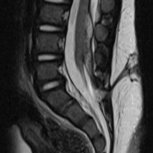
- If the tract is seen following birth, a magnetic resonance imaging (MRI) scan should be obtained. Images may show the tract and its point of attachment. MRI also shows masses within the canal.
Treatment
- Sinuses above the lumbosacral region should be surgically removed.
- Although approximately 25 percent of presumed sacral sinuses seen at birth will regress to a deep dimple on follow-up, all dermal sinuses should be surgically explored and treated prior to development of neurologic symptoms or signs of infection.
- The results of treatment following intradural infection are never as good as when undertaken prior to infection.
- Sinuses that terminate on the tip of the tailbone rarely penetrate the dura and may not need to be treated unless local infection occurs.


