


General Information
- A cavernous angioma is a blood vessel abnormality characterized by large, adjacent capillaries with little or no intervening brain. The blood flow through these vessels is slow.
- Cavernous angiomas can occur anywhere in the central nervous system.
- The disease occurs in 0.4 percent of the population, and 18.7 percent of these patients have multiple lesions.
Symptoms
- Symptoms include seizures, headache, hemorrhage or compression of surrounding brain tissue, called mass effect. Mass effect can cause weakness, numbness, double vision, visual disturbance or language difficulties.
- About 11 percent of lesions are not symptomatic.
Diagnosis
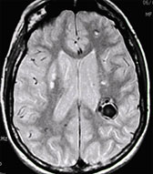
- Magnetic resonance imaging (MRI) is the most sensitive test for identifying cavernous angioma lesions. MRI scans often show small areas of new or old hemorrhages as a rim around the cavernous angioma. These lesions are not visible in cerebral angiography.

Treatment
- Patients with seizures are very likely to become symptomatic again. Seizure control frequently becomes more difficult with time, requiring surgical removal of the lesion.
- Surgical removal of the lesions offers the best possible outcome, but the risks of hemorrhage, age of the patient, presence of previous hemorrhages and the patient’s symptoms all must be considered.
- Symptomatic cavernous angiomas in children usually require treatment due to the high risk of future hemorrhage and greater seizure potential in children.
Outcome
- The consequences of a hemorrhage from a cavernous angioma are rarely catastrophic, in contrast with arteriovenous malformations (AVMs) or aneurysms.