


A colloid cyst is a slow-growing tumor typically found near the center of the brain.
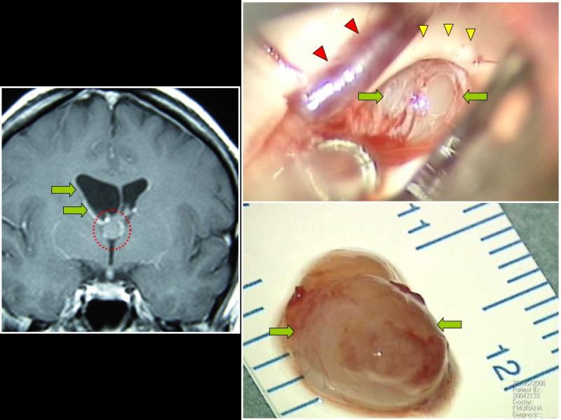
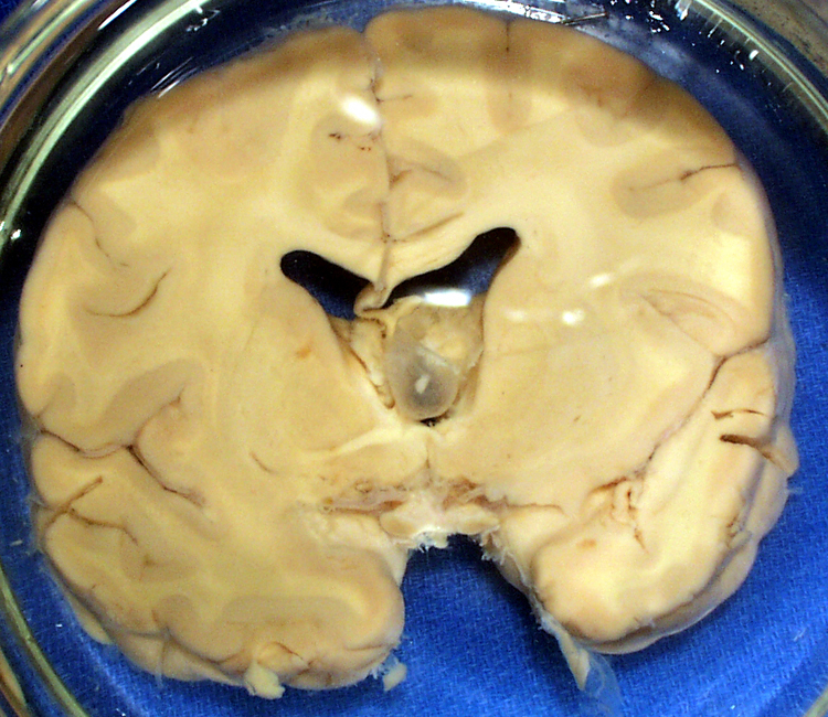
If large enough, a colloid cyst obstructs cerebrospinal fluid (CSF) movement, resulting in a build up of CSF in the ventricles of the brain (hydrocephalus) and elevated brain pressure.
Symptoms
- Most patients present with headaches, although other symptoms can occur.
- If symptomatic, prompt surgical treatment may be required in order to reduce the relatively high risk of sudden death.
Treatments
Minimally invasive surgery techniques
Minimally invasive techniques are used by us for the removal of colloid cysts. For patients with hydrocephalus, several options are available with us
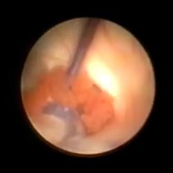
- Endoscopic removal – endoscopy typically requires very small bony opens.
Wehave expertise with both single and dual-port approaches
- 11.5-mm sleeve transcortical microsurgical removal
- The 11.5 mm diameter surgical port allows the use of a wider range of microsurgical instruments compared to the neuroendoscope.
- State-of-the-art image fusion and neuronavigation technology allow us to select the optimal pathway that minimizes brain tissue injury.
- The scalp incision size is similar to endoscopic technique
- Microcraniotomy interhemispheric microsurgical removal
- For patients with symptomatic or large colloid cysts but without significant hydrocephalus.
- Using state-of-the-art image fusion and neuronavigation technology, we can remove colloid cysts without the need to injure cortical brain tissue.
- We have expertise in surgical techniques that do not require the use of damaging brain retractors.
- The scalp incision size is only slightly longer than endoscopic and sleeve techniques
CSF shunt
- Placement of a shunt to drain CSF may be recommended if removal of the cyst is considered too high risk or if hydrocephalus continues despite removal of the cyst.